Introduction
Many individuals often ask, what is the difference between an ultrasound and sonogram, assuming both mean the same thing.While closely connected in medical practice, these terms describe different aspects of diagnostic imaging. Understanding their distinction can help patients make informed decisions and feel more confident during clinical procedures.
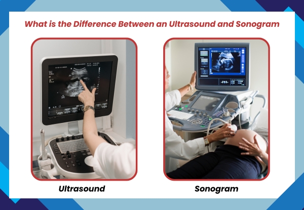
What Is an Ultrasound?

Ultrasound is a diagnostic imaging technique that uses high-frequency sound waves to capture live images from inside the body. It is commonly used in hospitals and clinics to assess organ function, detect abnormalities, and monitor fetal development.
How Ultrasound Imaging Works
A handheld device called a transducer sends sound waves into the body. These waves bounce off internal structures and return to the device, which then sends the data to a computer that forms an image. This procedure is non-invasive, safe, and does not involve radiation.
Common Uses of Ultrasound
Ultrasound is useful across multiple medical disciplines. Below are some frequent applications:
- Monitoring pregnancy and foetal health
- Examining liver, kidneys, and gallbladder
- Guiding needle placement for biopsies
- Checking for fluid buildup in joints or organs
- Evaluating blood flow using Doppler imaging
If you’re searching for a trusted facility, you may find options like an ultrasound clinic in Mumbai that offer comprehensive services tailored to both general and advanced diagnostic needs.
What Is a Sonogram?

A sonogram is the image produced by the ultrasound scan. While the ultrasound refers to the scanning technique, the sonogram is the visual result generated through that process. Healthcare professionals use this image to interpret what’s happening inside the body.
What Does a Sonogram Show?
Sonograms help doctors identify and monitor conditions such as cysts, organ enlargement, vascular blockages, or foetal positioning during pregnancy. While the terms are often used interchangeably, especially in conversations with patients, they serve different roles in the diagnostic process.
How It Differs from the Ultrasound Procedure
The distinction lies in their purpose: ultrasound is the method, while sonogram is the outcome. You could think of ultrasound as the action and sonogram as the result it produces.
Difference Between an Ultrasound and Sonogram
In clinical settings, the terms ultrasound and sonogram often overlap, but each has a specific role. Ultrasound is the medical procedure where sound waves are directed into the body to create images. It involves the use of a probe and computer to perform the scan. This scan does not emit radiation, making it safe for routine use, particularly in prenatal care and soft tissue evaluation.
Sonogram, on the other hand, refers to the static or moving images that result from the ultrasound scan. When a doctor reviews these images to diagnose a condition or track development, they are referring to the sonogram.
In daily practice, patients may hear either term used, depending on the context. A nurse might say you’re scheduled for an ultrasound, while the radiologist later discusses your sonogram results. The interchangeability in conversation doesn’t change the fact that they relate to distinct parts of the same process.
Understanding this distinction is especially important when choosing where to go for imaging. A patient searching for an ultrasound scan near me should expect both the procedure (ultrasound) and the result (sonogram) to be provided. Whether you’re consulting a best diagnostic center or a best ultrasound centre near me, knowing this difference allows you to ask the right questions and make informed decisions. Clinics like Midas Care Clinic offer clear explanations so patients feel supported and informed throughout their visit.
Types of Sonograms Used in Medical Settings
Different medical conditions require different sonographic techniques. These include:
- 2D Sonograms: Commonly used for general diagnostics and pregnancy imaging
- 3D and 4D Sonograms: Provide a more detailed view of foetal anatomy or structural anomalies
- Doppler Sonograms: Show blood flow through vessels, often used for cardiac and vascular assessments
- Transvaginal Sonograms: Offer a closer look at reproductive organs
Each of these types serves specific diagnostic goals and is chosen based on the patient’s symptoms or the doctor’s recommendations.
Common Situations Where Ultrasound and Sonograms Are Used
Pregnancy
Ultrasounds are widely used to monitor foetal growth, detect congenital abnormalities, and determine gestational age. The Midas Care Clinic and similar centres often recommend routine scans throughout pregnancy to ensure both mother and baby are healthy.
Abdomen and Pelvis
Doctors use abdominal ultrasounds to assess organs such as the liver, kidneys, gallbladder, and pancreas. Pelvic ultrasounds help evaluate reproductive organs and bladder conditions.
Muscles, Tendons, and Bones
Musculoskeletal ultrasound provides images of muscles, tendons, ligaments, and joints. It helps diagnose tears, inflammation, and other soft tissue concerns.
Blood Flow (Doppler Ultrasound)
Doppler techniques are used to evaluate circulation and detect vascular problems like blockages or reduced blood flow, especially in patients with cardiovascular conditions.
When Do Doctors Use the Term “Sonogram”?
Doctors and technicians sometimes use “sonogram” when referring to the images shared with patients. It’s more relatable and commonly used in discussions about pregnancy scans. However, medical documentation typically refers to the procedure as an ultrasound.
This variation in terminology does not affect the outcome or quality of care but highlights the importance of clear communication during diagnostic consultations.
5 uses of ultrasound in modern healthcare
Ultrasound technology is not limited to pregnancy. It plays an essential role in various medical disciplines. Here are five practical uses:
- Pregnancy monitoring: Assessing foetal development and detecting abnormalities.
- Abdominal scans: Evaluating liver, kidney, pancreas, and gallbladder conditions.
- Cardiac imaging: Analysing heart function using echocardiography.
- Musculoskeletal assessment: Imaging muscles, tendons, ligaments, and joints.
- Guidance for procedures: Supporting biopsies, injections, and catheter placements.
Choosing an ultrasound clinic in Mumbai
Selecting a reputable imaging facility ensures accurate diagnosis and timely reporting. In a large city like Mumbai, quality and trust are key.
Midas Care Clinic, recognised for its qualified professionals and patient care, offers a range of diagnostic services, including ultrasound scanning. Patients looking for reliable imaging can expect transparent processes and quick scheduling.
Frequently Asked Questions
Q1. What is the difference between an ultrasound and sonogram?
Answer: Ultrasound is the procedure using sound waves to scan inside the body. A sonogram is the image created from that scan.
Q2. Is a sonogram the same as an ultrasound?
Answer: No, a sonogram is the result of an ultrasound test.
Q3. Which term is used more in India?
Answer: Ultrasound is more common in medical usage, while sonogram appears in everyday conversations, especially around pregnancy.
Q4. Where can I get an ultrasound scan near me?
Answer: Most diagnostic centres and hospitals offer ultrasound services. Search locally or check with your physician.
Q5. How long does an ultrasound take?
Answer: Most scans take between 15 to 45 minutes, depending on the area being examined.
Q6. What are the main types of sonograms?
Answer: 2D, 3D, 4D and Doppler sonograms are the most commonly used types.
Q7. Do ultrasounds use radiation?
Answer: No, they rely on sound waves and are considered very safe.
Q8. Can ultrasounds guide treatment?
Answer: Yes. Ultrasound helps in guiding procedures like biopsies and catheter insertions.
Q9. How should I prepare for an ultrasound?
Answer: Preparation varies by test. Some may require fasting or drinking water before the scan.
Conclusion
Understanding the difference between an ultrasound and sonogram equips both patients and professionals with better clarity. One captures the process, the other the output. Whether you’re a medical practitioner or simply someone looking for answers, knowing this can improve communication and health outcomes.
Clinics such as Midas Care Clinic help bridge this understanding with precise imaging and expert consultation. Choosing the best diagnostic center ensures not just accurate results but also the peace of mind that comes with clarity.