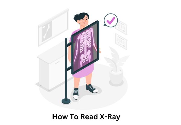
An X-ray is a type of imaging that allows doctors and healthcare professionals to look inside the body without making any cuts or incisions. Think of it as a snapshot of what’s happening beneath the skin, capturing detailed images of bones and soft tissues.
This non-invasive procedure is vital for diagnosing various conditions and injuries, making it one of the most common medical tests performed today.
In medical diagnostics, X-rays are widely used to examine broken bones, detect infections, or identify problems with organs like the lungs or heart. They can even help doctors assess dental health or screen for things like tumors. Given its ability to provide a clear view of the inside of the body, it plays a essential role in guiding treatment decisions and improving patient care.
As a patient or even someone just curious about the process, you may wonder: how to read an X-ray or understand the X-ray results. Knowing how to interpret an X-ray can help you get a better idea of what the doctor is looking for when they examine the images. In the following sections, we’ll explore simple steps to help you understand these essential results and make the process less intimidating.
What is an X-ray?
An X-ray is a special type of imaging test that uses radiation to create images of the inside of your body. It allows doctors to see bones, tissues, and organs without needing to make any incisions or cuts. In simpler terms, an X-ray acts like a camera that takes detailed pictures of what’s happening under your skin.
When an X-ray is taken, the radiation passes through your body, and different parts of your body absorb the radiation at different rates. For example, bones absorb more radiation and show up white on the image, while tissues like muscles or organs absorb less and appear darker. This difference in absorption creates the contrast needed to identify any issues.
There are several types of X-rays that serve different purposes:
- Chest X-rays: These are commonly used to check for lung conditions like pneumonia or tuberculosis, and they can also reveal heart problems.
- Bone X-rays: These are usually taken to check for fractures or bone-related issues. How to read X-rays of bones is essential for understanding fractures, cracks, or deformities.
- Dental X-rays: These help dentists see any hidden issues in the teeth and jawbones.
Understanding how an X-ray kaise dekhe (how to see an X-ray) can help you better understand what’s happening inside your body and make you more confident when reading your own X-ray report.
Why is It Important to Know How to Read X-Ray?
Knowing how to check X-ray report can be incredibly helpful, especially if you want to be more involved in your health decisions. Being able to understand the basic concepts of radiology tests and interpreting X-ray results can help you feel more informed when talking to your doctor about your health.
By learning how to read X-rays, you can get a better idea of what the X-ray images are showing, such as identifying areas of concern like fractures, lung issues, or infections. Additionally, having a basic understanding of radiology scan results can also empower you to ask the right questions and discuss potential treatment options with your healthcare provider.
Step-by-Step Guide on How to Read an X-Ray
Step 1: Understanding the Basics of X-ray Images
When you look at an X-ray, the first thing you’ll notice is the way the image shows different shades. This is due to the varying densities of tissues in your body.
- Bones: As we discussed, bones are denser, so they show up as white on the X-ray.
- Tissues and muscles: These appear darker because they absorb less radiation.
- Air spaces: Areas filled with air, such as the lungs, will show up as black or very dark.
This basic understanding of how to read X-rays of bones and how each density affects the image is essential when interpreting an X-ray report.
Step 2: Focus on Key Areas in X-ray Images
Once you understand the basic shades on an X-ray, the next step is focusing on the key areas. Common things to look for include:
- Lungs: When reading a chest X-ray, check for any unusual spots or shadows in the lungs. These might indicate problems like infections or fluid buildup.
- Heart: Ensure the heart’s size and shape are normal.
- Bones: Look for fractures or irregularities in bone structure, especially in bone X-rays.
Knowing how to read ultrasound reports can be helpful as well when comparing X-ray images to ultrasound diagnostics. These tests may work together to provide a fuller picture of your health.
Step 3: Identifying Abnormalities and Patterns
One of the most important aspects of reading an X-ray is learning how to spot abnormalities. Some things to look for include:
- Fractures: Cracks or breaks in the bones will appear as dark lines or breaks in the white areas.
- Infections: These might show as cloudy areas or abnormal shadows.
- Other issues: Tumors or foreign objects may also be detected.
Understanding these patterns will help you identify if something looks out of the ordinary in your X-ray reading and X-ray results.
Step 4: Understanding the Terminology in X-ray Reports
Your radiology report will often include terms that may be confusing at first. Here are some common terms and their meanings:
- Opacity: This refers to an area that is denser than the surrounding tissue, often indicating something abnormal, like an infection or tumor.
- Fractures: Broken bones will be noted as cracks or fractures.
- Density: The term refers to how much radiation is absorbed by an area, helping to indicate different types of tissue.
These terms are critical to understanding your radiology report and how to interpret the findings.
Common Mistakes When Reading X-Rays
Reading an X-ray might seem straightforward at first, but there are common mistakes people make when trying to interpret them. Here are a few errors to watch out for:
- Overlooking Small Details: Sometimes, minor fractures or abnormalities can be easy to miss if you’re not paying attention to the details. It’s important to carefully analyze every part of the X-ray.
- Misunderstanding Density Differences: Not understanding how different tissues absorb radiation can cause confusion. For example, soft tissues like muscles appear darker than bones, but if you don’t recognize this contrast, you might mistake an area for a problem.
- Relying on Personal Interpretation: While it’s helpful to understand how to read X-rays in simple terms, it’s important to remember that interpreting an X-ray kaise dekhe on your own may not always give the full picture. If you spot something odd, always seek professional help for an accurate diagnosis.
It’s essential to remember that reading X-rays requires training and experience. Relying too heavily on personal interpretation can lead to misunderstandings. If you’re unsure about something, consult a doctor or a radiologist.
How to Interpret X-ray Reports: A Quick Overview
Interpreting X-ray reports is a critical skill, but don’t worry it doesn’t have to be complex! Here’s a simplified way to approach it:
- Check the Report Details: First, ensure the report clearly identifies the area that’s been scanned. Is it a chest X-ray, a bone scan, or something else?
- Focus on the Key Findings: Look for any mentions of fractures, infections, or irregularities. For example, if your report mentions opacity, it could indicate an issue like a tumor or infection.
- Understand the Medical Terminology: Reports often use medical jargon like “density” or “fracture.” Familiarizing yourself with common terms like these will make X-ray report reading easier.
Common radiology tests like chest X-rays or bone X-rays often have their own sets of guidelines on how to interpret them. It’s always best to consult your doctor if you notice anything unusual.
Frequently Asked Questions
Q1: How do I read an X-ray report?
Ans: When you look at an X-ray report, focus on identifying the highlighted findings. Look for words like “fracture,” “opacity,” or “dense area.” Always take note of the area being scanned and the specific findings mentioned in the report. For instance, in a bone X-ray, fractures or cracks are the most common things to spot.
Q2: What does an X-ray report show?
Ans: An X-ray report shows images of the internal structures of the body. It helps doctors identify problems like fractures, infections, tumors, or issues with organs. For example, a chest X-ray helps detect lung conditions, while a bone X-ray shows fractures or bone-related issues.
Q3: Can I read an X-ray report myself?
Ans: You can try to understand your X-ray report yourself by looking for common terms like “fracture” or “opacity.” However, it’s important to remember that interpreting these images accurately requires medical expertise. It’s always best to consult a radiologist or doctor for the final interpretation.
Q4: What is the difference between a radiology scan and an X-ray?
Ans: A radiology scan is a general term that refers to any imaging technique used to see inside the body, including X-rays, CT scans, MRIs, and ultrasounds. An X-ray is just one type of radiology scan, specifically using radiation to capture images of bones and soft tissues.
Q5: How to identify a fracture in an X-ray?
Ans: Fractures in bone X-rays usually show up as dark lines or breaks in the white areas, which are the bones. These lines may vary in length and position, depending on the severity and type of fracture.
Q6: What does a chest X-ray show?
Ans: A chest X-ray provides images of the lungs, heart, ribs, and other chest organs. It’s commonly used to check for lung conditions, heart problems, or signs of infection.
Q7: Is it safe to read your own X-ray?
Ans: While it’s okay to try reading your own X-ray, it’s not recommended to make decisions based solely on your findings. It’s always safer to consult a doctor or radiologist to ensure that the interpretation is accurate and reliable.
Q8: What are the most common issues detected by X-rays?
Ans: Some of the most common issues detected by X-rays include:
- Fractures: Broken bones or cracks.
- Infections: Bacterial or viral infections in the lungs or bones.
- Tumors: Abnormal growths or masses in organs or bones.
Q9: How can I interpret an ultrasound report?
Ans: Interpreting an ultrasound report is similar to interpreting an X-ray report. Look for keywords related to findings in the report, like “normal” or “abnormal,” and ask your doctor to clarify any terms you’re unfamiliar with.
Q10: How does an X-ray differ from an MRI or CT scan?
Ans: An X-ray uses radiation to create images of bones and some soft tissues, while an MRI (Magnetic Resonance Imaging) uses magnets to produce detailed images of soft tissues, such as the brain or muscles.
A CT scan (Computed Tomography) combines multiple X-ray images to provide cross-sectional views of the body.
Conclusion
Understanding how to read X-rays is a valuable skill that can help you get a better grasp of your health, but always remember that a medical professional’s opinion is essential for an accurate diagnosis.
By focusing on the basics of X-ray results, understanding common terminology, and learning to identify key patterns, you can be more prepared when reviewing X-ray reports. However, it’s always recommended to seek expert advice for a thorough interpretation.
In case you need high-quality diagnostic services, consider visiting Midas Care Clinic, one of the best diagnostic centre in Mumbai, for reliable X-ray imaging and professional consultations.